Koi Disease Visual Field Guide
Educational guide · References and further reading below. Seek qualified advice for fish-health decisions.
Signs, diagnosis and first response for koi keepers | English | September 2026
Use this guide to match a change in behaviour or appearance with the right water test, skin examination or laboratory test. It covers 29 common diseases, visible syndromes and water emergencies in koi ponds and holding systems. Similar signs can have different causes, and more than one problem can occur at once.
If several koi are distressed: immediately increase aeration; stop feeding and medication; measure dissolved oxygen, ammonia, nitrite, pH, temperature and chlorine when relevant; record recent water changes, new arrivals and chemical doses. Stop fish and equipment movement if infection is possible.
Urgent professional help: gasping despite aeration, severe gill damage, rapid group deaths, deep ulcers, loss of balance, or possible KHV, CEV or SVC. Collect diagnostic samples before treatment when safe. Do not combine oxidisers or guess chemical doses.
Image key Real clinical koi and organism photographs sit beside their entries. Some microscope examples come from other fish or prepared specimens and are labelled. A photo cannot diagnose an individual koi. For external parasites, examine a fresh skin mucus sample promptly; leave gill sampling to a trained handler. Viruses require laboratory methods. Environmental injuries have no pathogen to image.
Choose the first check
| Pattern | First check | Why it matters |
|---|---|---|
| Several koi gasping or suddenly distressed | Oxygen, ammonia, nitrite, pH and chlorine after a water change | A shared water problem can kill rapidly and can resemble infection. |
| Rubbing, film, white specks or clamped fins | Water tests followed by a fresh skin mucus wet mount | Parasites and water irritation often look alike. |
| One enlarging red wound or ragged fin | Inspect injury, parasites and water; culture a progressive lesion | Treating bacteria alone may miss the initiating problem. |
| Rapid group illness after introducing koi | Stop movements; contact a fish veterinarian or diagnostic laboratory | KHV, CEV and SVC need laboratory tests and biosecurity. |
Reading each entry Cause explains the mechanism; Early warning is what to spot first; Developed signs describe progression; Confirm tells you the useful test; Response gives immediate steps and treatment direction; Distinguish highlights a common mistake or follow-up.
External parasites and water mould
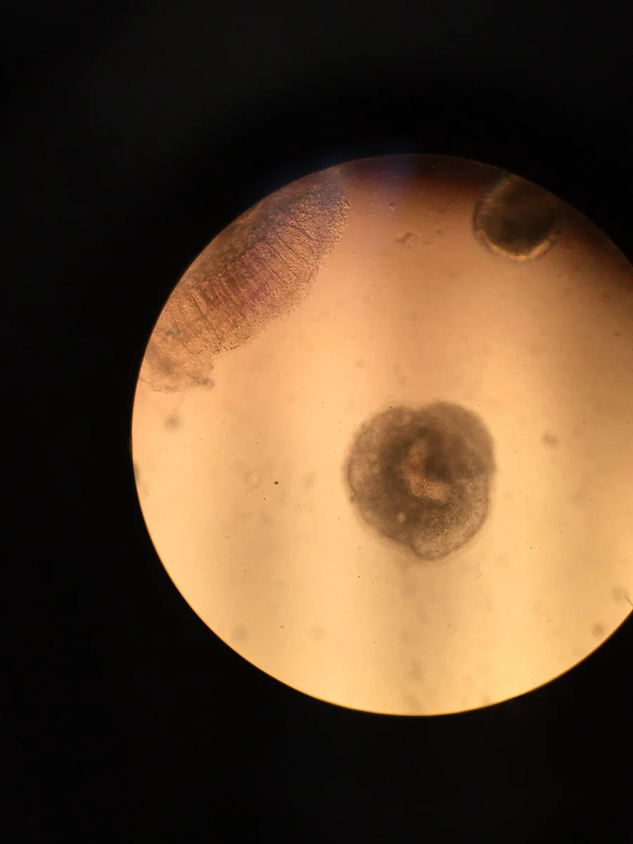
Ich [M1]. USFWS Mountain Prairie, public domain. Source and licence https://commons.wikimedia.org/wiki/File:Parasite_Under_a_Microscope_(51297061033).jpg
White spot Ich Cause: A ciliate that passes through attached, encysted and free-swimming stages. Early warning: Flashing, fast breathing; white grains may appear later. Developed signs: White pinhead grains may spread to fins and skin; heavily affected gills may fail before spots are obvious. Confirm: Fresh skin or gill wet mount; large rotating ciliate [M1]. Response and treatment: Aerate, check water, then use a labelled koi anti-protozoal course covering repeated free-swimming stages. Distinguish or prevent: Repeat treatment must follow the parasite life cycle and product label; an isolated single bath often misses protected stages. View affected koi photograph https://koiorganisationinternational.org/blog-entry/why-do-my-koi-have-white-spots
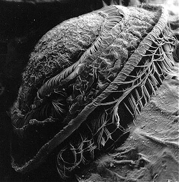
Trichodina [M2]. Mullet specimen. A D M Dove, attribution required. Source and licence https://commons.wikimedia.org/wiki/File:Trich2.jpg
Trichodina Cause: A disc-shaped ciliate that proliferates with organic waste and stressed fish. Early warning: Grey film, excess mucus, rubbing, clamped fins. Developed signs: Mucus thickens and fish may darken or hold their fins close to the body. Confirm: Wet mount: spinning disc with toothed ring [M2]. Response and treatment: Reduce organic waste; treat confirmed heavy burdens with an appropriate labelled anti-protozoal. Distinguish or prevent: A few organisms can occur without disease; interpret the burden with fish condition and water tests.
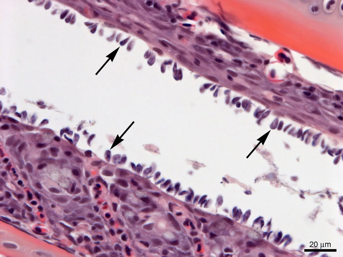
Ichthyobodo [M3]. Gill section. Carla M Conway, public domain. Source and licence https://commons.wikimedia.org/wiki/File:Ichthyobodo_gill_infection.jpg
Ichthyobodo Costia Cause: A very small flagellate on the skin and gills, sometimes associated with rapid losses. Early warning: Fine grey haze, clamping, rapid gill distress. Developed signs: A dull bluish-grey film and laboured breathing can progress quickly, especially in crowded systems. Confirm: Examine a fresh wet mount promptly at higher power [M3 is histology]. Response and treatment: Urgent oxygen and water correction; arrange a koi-safe anti-protozoal treatment after confirmation. Distinguish or prevent: Examine a fresh, promptly prepared sample; delayed slides make this mobile organism harder to find.
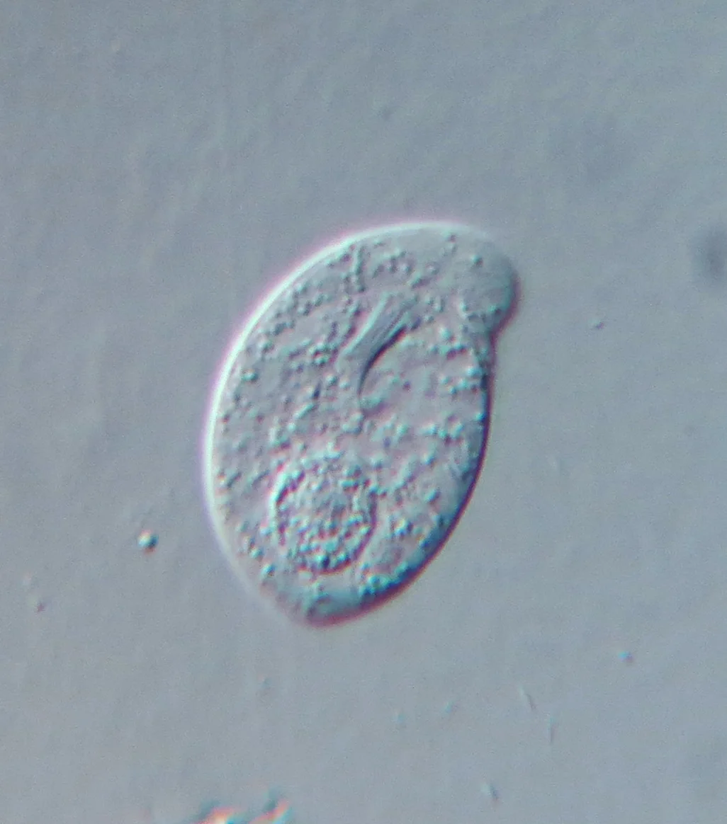
Chilodonella [M4]. Picturepest, CC BY 2.0. Source and licence https://commons.wikimedia.org/wiki/File:Chilodonella_uncinata_-_400x_(8999778877).jpg
Chilodonella Cause: A flattened skin or gill ciliate that may multiply rapidly in stressed fish. Early warning: Pale film, mucus, flashing; fast breathing in advanced cases. Developed signs: Excess mucus, pale skin and rising respiratory effort may follow initial rubbing. Confirm: Wet mount: flattened gliding ciliate [M4]. Response and treatment: Increase aeration; treat a confirmed infection with a suitable labelled protocol. Distinguish or prevent: The film resembles Costia, Trichodina and water irritation; confirm the moving organism before selecting treatment.
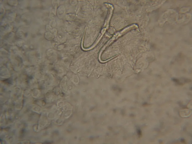
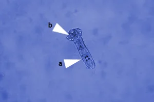
M5: Maxikoi, CC BY-SA 3.0. Image source https://commons.wikimedia.org/wiki/File:Gyrodactylus_sp.jpg M6: Gogoschinski, public domain. Image source https://commons.wikimedia.org/wiki/File:Dactylogyrus-Mikroskop.jpg
Skin and gill flukes Cause: Monogenean parasites attach by hooks; skin forms often bear live young, while many gill forms lay eggs. Early warning: Flashing, yawning, one gill cover partly closed, breathing effort. Developed signs: Persistent rubbing, mucus and one-sided gill irritation may become severe respiratory distress. Confirm: Skin and gill wet mounts; identify hooks, eggs or embryos [M5 M6]. Response and treatment: Praziquantel is often used; plan repeat checks or treatment for egg-laying gill flukes. Distinguish or prevent: Eggs can survive an initial treatment; plan follow-up microscopy and a product-appropriate repeat schedule.
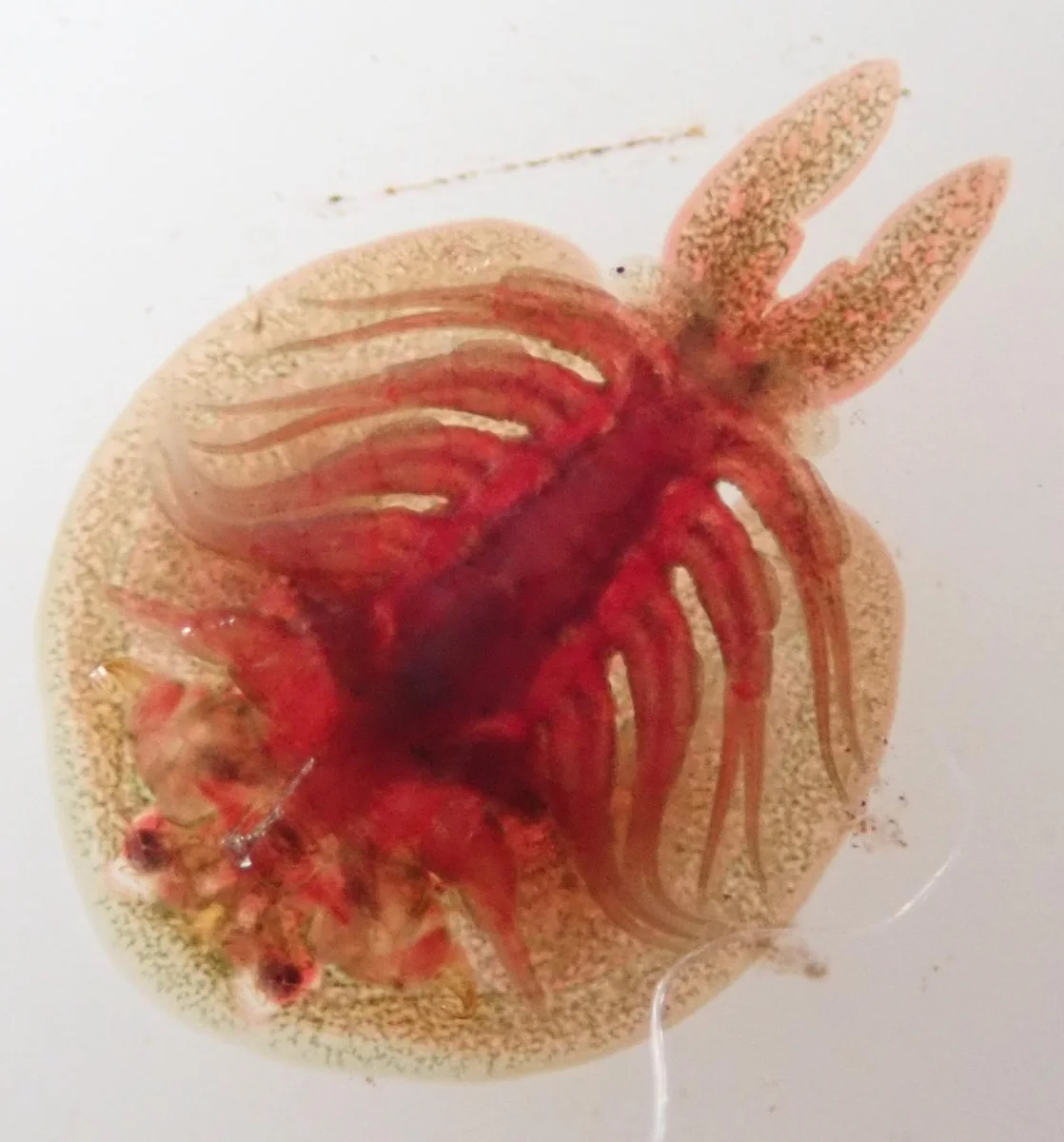
Fish louse Argulus [M7]. Robert Taylor, CC BY 4.0. Source and licence https://commons.wikimedia.org/wiki/File:Genus_Argulus_Fish_Louse_Rob_Taylor.jpg
Fish lice Argulus Cause: A mobile crustacean parasite that feeds on koi and lays eggs away from the fish. Early warning: Violent flashing, jumping, moving discs and small red punctures. Developed signs: Small haemorrhagic punctures and ulcers can appear after intense scratching and jumping. Confirm: Visual inspection or magnification [M7]. Response and treatment: Treat fish and life stages in the system; inspect wounds. Manual removal alone does not clear eggs. Distinguish or prevent: Check pond surfaces and new arrivals; removing visible adults does not remove environmental eggs.
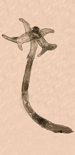
Anchor worm Lernaea [M8]. US Geological Survey, public domain. Source and licence https://commons.wikimedia.org/wiki/File:Lernaea_cyprinacea.jpg
Anchor worm Lernaea Cause: A copepod whose adult female embeds an anchor-like head under the skin. Early warning: A protruding thread with local redness and scale lifting. Developed signs: The projecting thread can leave a deep inflamed wound that develops secondary infection. Confirm: Inspect attachment and paired egg sacs [M8]. Response and treatment: Remove adults only with skilled handling; manage free stages and secondary wounds. Distinguish or prevent: Avoid tearing tissue during removal; free-swimming stages remain in the system.
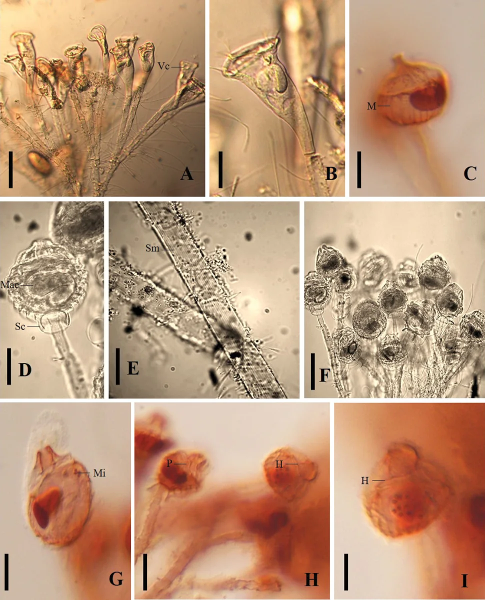
Epistylis [M9]. Ramírez-Ballesteros et al., CC BY 4.0. Source and licence https://commons.wikimedia.org/wiki/File:Epistylis_hentscheli_(10.3897-zookeys.782.26417)_Figure_2.jpg
Epistylis Cause: A stalked colonial ciliate that may grow on damaged skin and organic debris. Early warning: Grey-white focal tufts with irritation or a wound underneath. Developed signs: Small grey-white clusters can resemble fungus, often over a primary injury or ulcer. Confirm: Wet mount: branching stalks with bell-shaped zooids [M9]. Response and treatment: Reduce organic load; investigate underlying tissue damage; treat only after identification. Distinguish or prevent: Treat the underlying injury and water conditions; a visible tuft does not prove Epistylis is the primary cause.
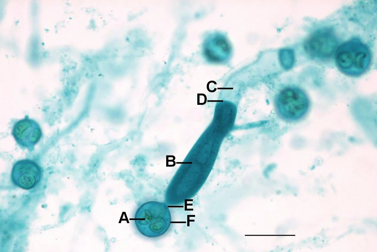
Saprolegnia [M10]. Jon Houseman, CC BY-SA 3.0. Source and licence https://commons.wikimedia.org/wiki/File:Oomy1000L.jpg
Saprolegnia water mould Cause: An oomycete that commonly colonises injured tissue and dead eggs. Early warning: Cotton-like growth on a wound, damaged fin or dead egg. Developed signs: Cotton-like tufts enlarge over an abrasion, fin tip or egg mass. Confirm: Wet mount: broad branching hyphae [M10]. Response and treatment: Correct the original injury; use an appropriate labelled water-mould product when needed. Distinguish or prevent: Unlike columnaris, this usually has broad branching hyphae on microscopy; find the original injury.
Bacterial disease and visible syndromes
Other conditions in this group These entries have no embedded diagnostic photograph. Use the listed test before treatment.
| Recognise | Confirm and act |
|---|---|
| Bacterial ulcer Cause A progressive skin wound often complicated by Aeromonas or other opportunistic bacteria. Early One red point or lifted scale progresses to an open ulcer. Progression A red pinpoint or raised scale can become an open crater with tissue loss. View koi ulcer photo https://www.merckvetmanual.com/multimedia/image/hemorrhagic-ulcer-body-wall-koi | Confirm Scrape for parasites; culture a progressive lesion before antibiotics. Response Improve water and oxygen; fish veterinarian for wound care and culture-guided systemic treatment. Watch Water problems, handling injury and parasites can start the lesion; correct these while arranging culture. |
| Columnaris mouth or skin disease Cause A Flavobacterium infection that can affect mouth, skin, fins and gills. Early Pale mouth rim, saddle-like patch, rapid fin or gill injury. Progression A pale mouth rim or saddle-shaped patch can progress to rapid necrosis and breathing distress. | Confirm Bacterial examination and culture; cottony appearance can mimic mould. Response Reduce crowding and stress; seek prompt professional antibacterial treatment. Watch Cottony appearance is not proof of water mould; gill involvement makes the case urgent. |
| Fin erosion Cause A visible syndrome caused by injury, poor water, parasites or bacterial invasion. Early Opaque or red fin edge, progressive fraying. Progression The fin edge may turn opaque, red or ragged before shortening. | Confirm Check sharp surfaces, water, parasites; culture advancing lesions. Response Remove cause; refer deep or advancing tissue loss for culture-guided treatment. Watch Check other fish and sharp fittings; distinguish an isolated tear from progressive tissue loss. |
| Bacterial gill disease Cause Bacterial overgrowth on stressed gills, often alongside crowding or water problems. Early Faster breathing, gathering at returns; sometimes no skin lesion. Progression Rapid breathing may advance without an obvious skin lesion; fish seek high-flow areas. | Confirm Oxygen and water panel, gill wet mount, bacterial evaluation. Response Maximum aeration and clean water; professional diagnosis before gill-sensitive medication. Watch Oxygen shortage, ammonia, chlorine and flukes look similar; test water before using gill-irritating chemicals. |
| Dropsy or septicaemia Cause A syndrome of fluid accumulation or systemic illness with several possible causes. Early Reduced appetite, swelling, raised scales or protruding eyes. Progression Swelling, protruding eyes and eventually pinecone scales may accompany loss of appetite. | Confirm Exam, imaging or culture; dropsy is a sign with several causes. Response Isolate safely in excellent water; urgent veterinary assessment. Prognosis worsens with pineconing. Watch Pineconing is a late warning sign; no single antibiotic or salt dose cures every underlying cause. |
| Mycobacterial disease Cause A chronic infection caused by aquatic mycobacteria that may also infect broken human skin. Early Long-term wasting, nodules, deformity or chronic ulcers. Progression Fish may waste slowly, develop nodules, spinal changes or non-healing ulcers. | Confirm Laboratory histology, acid-fast stain and culture; ordinary wet mount is insufficient. Response Stop stock movement and seek a diagnostic plan; wear gloves over broken skin. Watch Use gloves for wounds, dedicated equipment and laboratory diagnosis; indiscriminate antibiotics rarely solve a facility problem. |
| Popeye Cause Exophthalmia is a sign of injury, gas or internal disease rather than one diagnosis. Early One eye protrudes after injury; both eyes can indicate systemic trouble. Progression One eye may cloud or protrude after trauma; both eyes plus swelling suggests a broader problem. | Confirm Check trauma, water, gas bubbles and body swelling. Response Remove injury source; severe, bilateral or progressive cases need a fish veterinarian. Watch Check for a sharp impact point, total gas problems and systemic disease before deciding on treatment. |
| Hikui and persistent red lesions Cause A persistent erosive lesion associated with red pigment in koi; other causes can mimic it. Early Slow rough or eroded patch within red pigment, often recurrent. Progression A rough red patch may recur or slowly enlarge without the pattern of an acute ulcer. | Confirm Experienced exam, sometimes biopsy; exclude bacterial ulcer and tumour. Response Document growth under consistent light and obtain specialist assessment before cautery. Watch Photograph changes, seek specialist examination and exclude infection or tumour before cautery. |
Viral disease and biosecurity
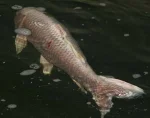
KHV affected koi [C1]. Jdimmers, CC BY-SA 3.0. Source and licence https://commons.wikimedia.org/wiki/File:Khvinfectedkoi.png
Koi herpesvirus KHV Cause: A contagious carp herpesvirus that may leave surviving koi as carriers. Early warning: Group lethargy, appetite loss, gill distress after introduction [C1]. Developed signs: At permissive temperatures, appetite loss and lethargy can progress to pale, patchy gills and group deaths. Confirm: Laboratory PCR on correctly collected tissue; wet mount cannot confirm. Response and treatment: Stop all fish and equipment movement; oxygen support and urgent laboratory or fisheries advice. No antiviral cure. Distinguish or prevent: Quarantine and diagnostic PCR are essential; changing water temperature is not a cure or a reliable way to clear carriers.
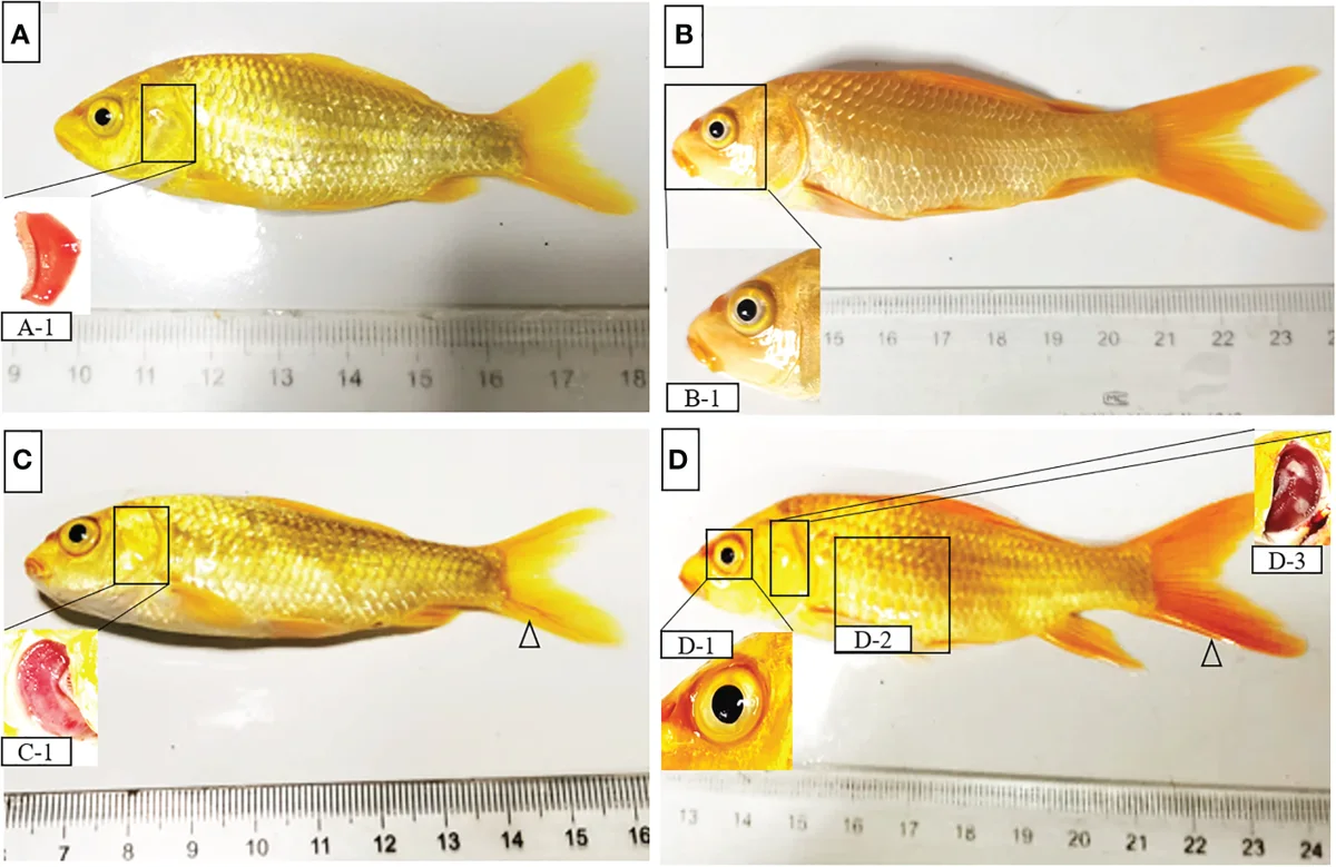
CEV affected koi [C2]. Study series includes controls. Ouyang et al., CC BY 4.0. Source and licence https://doi.org/10.3389/fimmu.2023.1142830
Carp edema virus CEV Cause: A poxvirus associated with koi sleepy disease and gill damage. Early warning: Bottom sitting or sleepy behaviour, gill distress [C2]. Developed signs: Bottom sitting, extreme lethargy and slow gill movements can lead to sudden losses. Confirm: Validated laboratory PCR; check for concurrent parasites. Response and treatment: Isolate equipment and stock; maintain oxygen and stable water. No specific antiviral cure. Distinguish or prevent: Normal-looking koi can share exposure; use PCR and maintain strict separation of stock and equipment.
Other conditions in this group These entries have no embedded diagnostic photograph. Use the listed test before treatment.
| Recognise | Confirm and act |
|---|---|
| Spring viraemia of carp SVC Cause A systemic viral infection of carp that tends to present in cooler water. Early Lethargy, haemorrhage, swelling and deaths, often in cooler water. Progression Lethargy, bleeding, bulging eyes, swelling and group deaths can resemble bacterial septicaemia. View confirmed koi photo https://www.usgs.gov/media/images/investigation-and-disease-prevention-spring-viremia-carp-virus | Confirm Official laboratory testing; signs overlap bacterial septicaemia. Response Stop movements and contact fish-health or fisheries authority. No specific curative treatment. Watch Do not rely on appearance or sell or move exposed fish while a laboratory and fish-health authority investigate. |
| Carp pox CyHV 1 Cause A different carp herpesvirus that produces benign-looking skin overgrowths in older koi. Early Smooth waxy plaques, often with normal appetite. Progression Smooth, waxy white or grey plaques on fins or skin are often more obvious in cooler periods. View koi pox photo https://www.tiho-hannover.de/kliniken-institute/fachgebiete-und-forschungszentren/abteilung-fischkrankheiten-und-fischhaltung/forschung-und-projekte/virusinfektionen/karpfenpocken-virus-cyprinides-herpesvirus-1 | Confirm Clinical exam; biopsy when lesion is atypical. Response Monitor with excellent husbandry; avoid cutting lesions. Virus may persist. Watch Do not cut off plaques; ask for biopsy if a lesion is ulcerated, rapidly growing or atypical. |
Water and environmental emergencies
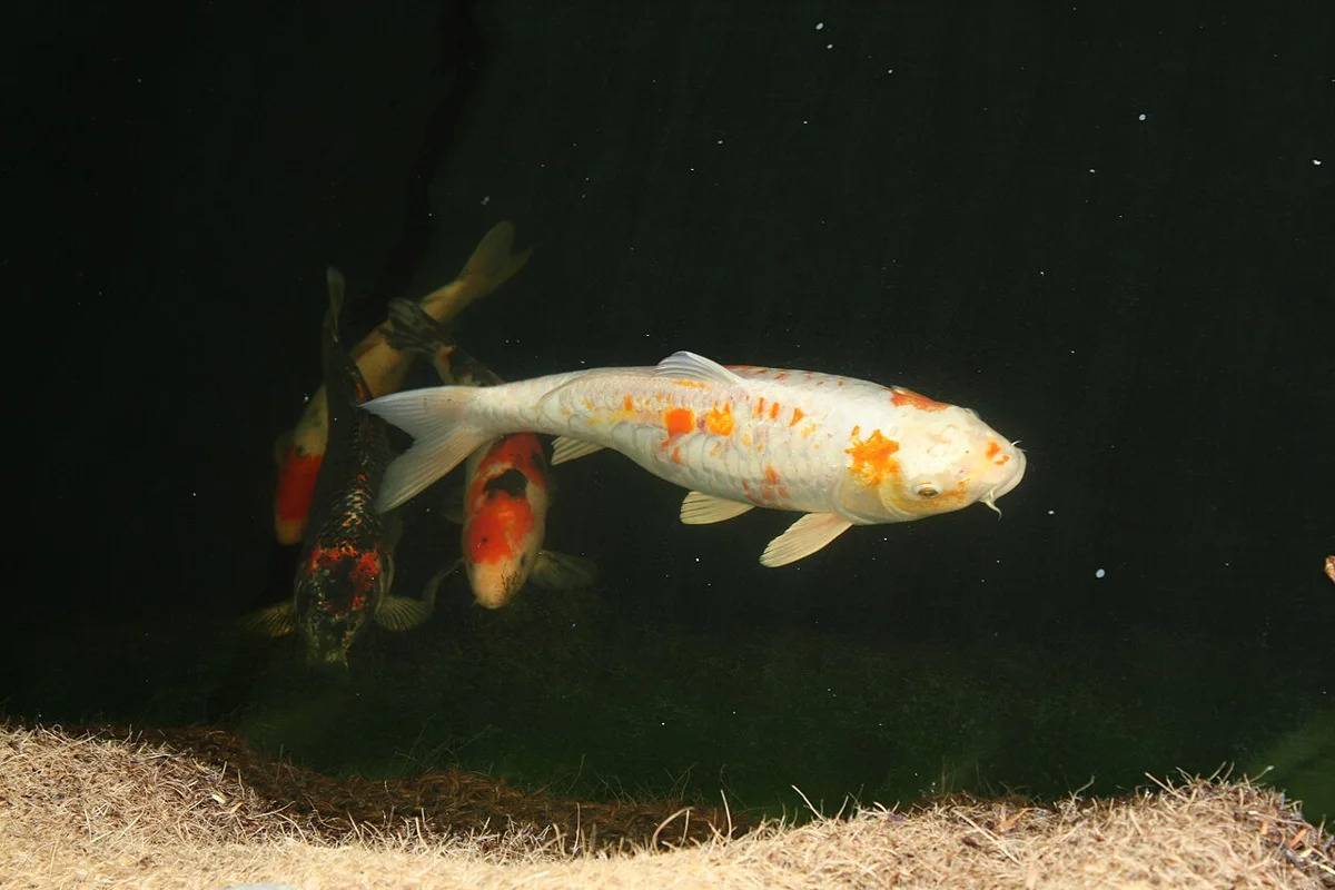
Koi with sunburn [C3]. TaylorNews, CC BY-SA 3.0. Source and licence https://commons.wikimedia.org/wiki/File:Koi_with_sunburn.JPG
Sunburn or handling trauma Cause: UV exposure or netting and transport can injure koi skin. Early warning: Red veins, abrasions or lost scales after exposure [C3]. Developed signs: Raw areas may widen or become secondarily infected. Confirm: Check the exposure history and water; scrape persistent lesions. Response and treatment: Shade, aerate and maintain clean water; handle gently and reassess. Distinguish or prevent: Spreading redness or several ill fish need an infection and water work-up.
Other conditions in this group These entries have no embedded diagnostic photograph. Use the listed test before treatment.
| Recognise | Confirm and act |
|---|---|
| Ammonia toxicity Cause Un-ionised ammonia damages gills; its toxic fraction rises as pH and temperature rise. Early Multiple fish restless, red gills or gasping after filter trouble. Progression All fish may become restless, gasp or show red gills after overfeeding or filter failure. | Confirm Total ammonia plus pH and temperature; assess toxic NH3 fraction. Response Stop feed, aerate, controlled dechlorinated changes, protect biofilter and retest. Watch A clear pond can still contain toxic ammonia; record total ammonia, pH and temperature together. |
| Nitrite toxicity Cause Nitrite interferes with oxygen transport after biofilter disruption; chloride reduces uptake at the gill. Early Fast breathing with high nitrite; brown blood is a later clue. Progression Fish breathe rapidly even when oxygen is adequate; brown blood is a late sign. | Confirm Nitrite test with units checked; assess chloride and oxygen. Response Stop feed, restore filtration; calculated chloride support may help under expert direction. Watch Check test units and chloride before calculating any salt response; restore biofiltration. |
| Low dissolved oxygen Cause Oxygen falls with poor circulation, heavy biomass, plant respiration or equipment failure. Early Fish gather at waterfall or surface, especially near dawn. Progression Multiple koi gather near air or waterfall outlets, often worst before dawn. | Confirm Measure dissolved oxygen during distress, including at depth. Response Immediate vigorous aeration and circulation; stop feeding and oxygen-consuming chemicals. Watch A single daytime reading may miss the trough; measure during distress and restore aeration immediately. |
| Chlorine or chloramine injury Cause Untreated municipal water or an inadequate conditioner damages gills. Early Sudden distress during or after a tap-water change. Progression Fish may flash, gasp or die soon after a water change while source water looks normal. | Confirm Free and total chlorine test; check conditioner and source volume. Response Stop inflow, use an appropriate full-volume conditioner, aerate and monitor ammonia. Watch Chloramine can leave ammonia after neutralisation; test both disinfectant and ammonia after correction. |
| pH crash or rapid swing Cause Low alkalinity or abrupt dosing lets pH change enough to stress gills and skin. Early Whole-pond flashing, clamping or gasping after rain or dosing. Progression A whole pond may clamp, rub or gasp after rain, a large change or chemical treatment. | Confirm Morning and afternoon pH, KH, ammonia and temperature. Response Aerate, stop feed, correct buffering gradually after testing ammonia. Watch Measure KH and pH over time; sudden pH correction can increase toxic un-ionised ammonia. |
| Gas bubble disease Cause Excess dissolved gas can form bubbles within tissues, often after pressurised water or pump leaks. Early True bubbles in fin, skin or eye tissue near a return. Progression Fine bubbles in fins, skin or eyes and erratic behaviour may follow water-system changes. | Confirm Measure total gas pressure; inspect suction-side leaks and water source. Response Stop the supersaturated source and degas water; seek help for severely affected fish. Watch Surface bubbles alone are not diagnostic; check total gas pressure and plumbing. |
| Buoyancy or abdominal swelling Cause A sign that may arise from internal disease, injury, gas, masses or fluid. Early Tilting, rolling, sinking or asymmetric abdomen. Progression Tilting, rolling or sinking can be accompanied by an uneven abdomen or reduced feeding. | Confirm Exam and imaging when persistent; not every case involves a swim bladder. Response Maintain good water and prevent abrasions; obtain a cause-specific veterinary plan. Watch Avoid treating every floating fish as a swim-bladder infection; persistent signs need examination and imaging. |
Keep a useful case record
For a valuable koi or a group outbreak, record these details before you change water or start treatment when it is safe to do so. They help a fish veterinarian distinguish a pond problem from a contagious one.
| Record | Why it helps |
|---|---|
| Pond and fish history | Pond or tank ID, number and sizes of affected koi, new arrivals, mixing, feeding and first day of abnormal behaviour. |
| Water at the time | Dissolved oxygen, temperature, pH, KH, total ammonia, nitrite and chlorine after municipal water; include units and test time. |
| Images and observations | Clear whole-fish and close-up photos, breathing rate, rubbing, appetite, gill appearance and daily deaths. |
| Samples and treatments | Ask the receiving laboratory how to submit live sick fish or tissue; record prior medicines and doses. A recent chemical treatment can alter tests. |
References and further reading
This guide follows veterinary and university fish-health references. Confirm the diagnosis, pond volume and water conditions before following a current local treatment label. A fish veterinarian should direct systemic antibiotics.
Merck Parasites | Merck Bacteria | Merck Viruses | UF IFAS KHV https://www.merckvetmanual.com/exotic-and-laboratory-animals/aquarium-fish/parasitic-diseases-of-fish https://www.merckvetmanual.com/exotic-and-laboratory-animals/aquarium-fish/bacterial-diseases-of-fish https://www.merckvetmanual.com/exotic-and-laboratory-animals/aquarium-fish/viral-diseases-of-fish https://ask.ifas.ufl.edu/publication/VM113
UF IFAS CEV | UF IFAS Fish Health | UF IFAS Ich | UF IFAS Flukes https://ask.ifas.ufl.edu/publication/fa189 https://ask.ifas.ufl.edu/publication/FA004 https://ask.ifas.ufl.edu/publication/FA006 https://ask.ifas.ufl.edu/publication/FA033
UF IFAS SVC | UF IFAS Ammonia | UF IFAS Salt and Nitrite https://ask.ifas.ufl.edu/publication/VM106 https://ask.ifas.ufl.edu/publication/FA031 https://ask.ifas.ufl.edu/publication/VM007