Red Veins and Reddening in Koi
Educational guide · References and further reading below. Seek qualified advice for fish-health decisions.
How to distinguish stress and irritation from injury water quality problems parasites and systemic disease
Red veins or a pink flush can appear suddenly on a white-skinned koi and create immediate fear that the fish has a bacterial infection. Sometimes that fear is justified. External reddening and haemorrhage are recognised features of haemorrhagic septicaemia in fish. Yet the same broad visual complaint can also follow transport, rough netting, spawning, low dissolved oxygen, ammonia, chlorine exposure, an unstable pH, parasites, a local wound or another source of inflammation. Redness is therefore an important sign, but it is not a diagnosis.
The safest response begins by asking what pattern is present, how quickly it developed, whether one fish or several are affected, and what changed in the pond before the colour appeared. A few visible vessels in a translucent tail are not equivalent to scattered pinpoint bleeding across the body. A local red patch after a collision is not equivalent to diffuse reddening accompanied by ulcers, swelling and rapid deterioration. The colour must be interpreted together with breathing, behaviour, appetite, water results and the recent history.
This article explains what red veins and red skin may represent, how to assess urgency, what to test during the first hour, when microscopy or bacterial culture is useful, and why treatment should target the cause rather than the colour. It is written for koi keepers and dealers managing outdoor ponds, quarantine vats, sales systems and transport-related cases, including warm tropical conditions.
Quick answer
Red veins and reddening in koi may reflect increased visibility of superficial blood vessels, active vasodilation, local inflammation, tissue injury or true haemorrhage. Common triggers include handling and transport stress, warm water and low oxygen, ammonia or chlorine injury, rapid pH change, parasites, abrasion, developing ulcers and systemic bacterial disease. Appearance alone cannot tell these apart.
Treat the case as urgent when reddening is spreading rapidly, more than one koi is affected, breathing is abnormal, pinpoint haemorrhages or fin-base bleeding are present, or the fish also shows ulcers, pop-eye, swelling, pineconing, loss of balance, severe lethargy or deaths. Immediately improve reliable aeration, stop unnecessary handling, test the actual water and review recent events. Do not add antibiotics, oxidisers, antiparasitic treatments or salt simply because the fish looks red.
The central rule is simple: a red koi should trigger an investigation before it triggers a medication. Correct a measured environmental problem immediately. Use a fresh skin or gill examination when parasites are plausible. Seek fish-health or laboratory support when systemic disease, multiple affected fish or unexplained mortality is present.
How to read redness as a clinical sign
The visible surface of a koi includes scales, epidermis, mucus, pigment cells and a network of small blood vessels beneath or within the tissues. White areas provide little pigment to hide that network, so vascular change is often easiest to see on shiroji, the pale fins and the tail. The apparent intensity also changes with lighting, viewing angle, water clarity and whether the fish is seen through water or in a bowl.
Redness can be produced in several different ways. Hyperaemia means more blood is actively flowing through a tissue, usually because vessels have widened during heat, irritation or inflammation. Congestion describes blood accumulating because drainage from an area is impaired. Haemorrhage means blood has escaped from vessels into surrounding tissue. These processes can look similar from a distance, but their implications differ.
Fine lines that follow fin rays or a small number of superficial vessels can become more obvious during stress without obvious bleeding into the tissue. A diffuse blush is broader and less sharply defined. Petechiae are small pinpoint haemorrhages. Ecchymoses are larger irregular areas of bleeding. Red streaks at the fin base, around the vent, beneath scales or across the body are more concerning when they are new, widespread or accompanied by other systemic signs.
Do not judge from colour alone. A white koi under a warm light can look pinker than it did under daylight. A recently bowled fish may show more obvious vessels because capture and crowding increased activity and stress. Compare the fish with a photograph taken under similar conditions, then look for functional changes such as breathing, posture and appetite.
The main visual patterns
| Pattern | What it may represent | What makes it more concerning |
|---|---|---|
| Fine red lines in translucent fins | Visible or dilated vessels; stress, heat, handling or early irritation may contribute | New rapid increase, several fins affected, clamping, breathing change or red streaking at the fin bases |
| Diffuse pink blush on white skin | Vasodilation, environmental irritation, chemical exposure or inflammation | Several fish change together, recent water change or treatment, fast breathing, mucus change or worsening over hours |
| One local red patch | Collision, net injury, spawning abrasion, parasite attachment or early ulcer | Scale loss, swelling, heat-like raised tissue, discharge, deepening centre or rapid expansion |
| Pinpoint red dots | Petechial haemorrhage caused by vessel injury, toxin exposure or systemic disease | Widespread distribution, fin-base bleeding, lethargy, swelling, ulcers or deaths |
| Red streaks in fins and body | Inflammation or haemorrhage; can occur in bacterial septicaemia but is not specific | Progression despite good water, multiple systems affected, anorexia, exophthalmia or abnormal swimming |
| Red or inflamed gills | Gill irritation, ammonia, chlorine, parasites, bacterial disease or physical damage | Gasping, surface piping, pale or mottled areas, necrosis, sudden deaths or several fish involved |
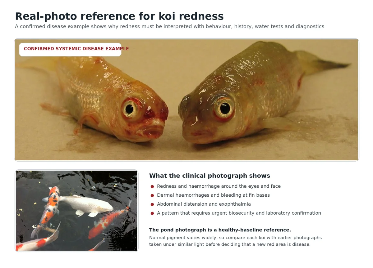
Figure 1 Real-photo reference. Top: koi with confirmed spring viraemia of carp virus showing dermal haemorrhages, redness around the eyes, abdominal distension and bleeding at fin bases. This is one confirmed cause of severe redness, not a visual diagnosis for every red koi. Bottom: healthy pond koi illustrating the value of a normal baseline. Clinical photograph by Eveline Emmenegger, USGS, public domain. Pond photograph by Laslovarga, Wikimedia Commons, CC BY-SA 3.0, cropped.
Why white koi can look suddenly red
Redness is often noticed first on Kohaku, Sanke, Showa, Shiro Utsuri and other koi with large pale areas because the skin provides contrast. That does not mean white varieties are uniquely affected by every cause. The same vascular or inflammatory event may be less visible on a dark, metallic or heavily patterned fish.
Skin quality and thickness, scale type, age, condition and recent growth can influence visibility. Doitsu koi may show changes differently because much of the body lacks the regular layer of scales found in Wagoi. Thin fins reveal vessels more readily than the trunk. A judgement based on a single photograph should therefore account for variety and anatomy, but these factors must not be used to dismiss a genuinely new change.
A practical baseline is valuable. Photograph valuable koi when they are healthy, using a consistent bowl, daylight or fixed light, and a top-down angle. Record temperature and recent handling. Later comparisons are far more reliable than asking whether the fish looks red in an isolated image.
Environmental and chemical causes
Low dissolved oxygen and heat
Warm water contains less dissolved oxygen than cool water, while koi, nitrifying microorganisms and decomposing organic material continue to consume it. In a heavily stocked pond or holding vat, oxygen may fall before sunrise, after a pump failure, during a treatment, following a heavy feeding event or when algae and bacteria consume oxygen at night. Fish may increase gill movement, crowd returns, hang near the surface or become restless.
Heat and exertion can make vessels more visible, but low oxygen should never be diagnosed from redness alone. Measure dissolved oxygen if possible and observe the whole population. Large koi may show distress first because their oxygen demand is high. Immediate aeration is appropriate when breathing is abnormal even before every test result is available.
An aeration response does not prove oxygen was the only cause. A koi with gill parasites or systemic infection can also seek high-flow water. The useful interpretation is that oxygen support buys safety while the investigation continues.
Ammonia and biofilter failure
Ammonia arises from fish metabolism, uneaten feed and decomposing organic matter. A new or damaged biofilter, sudden increase in feeding, dead fish, blocked mechanical filtration, medication damage to nitrifying organisms or loss of circulation can allow total ammonia nitrogen to rise. The more toxic un-ionised ammonia fraction increases as pH and temperature rise, so a total-ammonia reading must be interpreted with those measurements.
Ammonia injures gill and other epithelial tissues and can produce abnormal breathing, excess mucus, irritation, lethargy and colour change. Red or inflamed gills may be present, while the external skin may appear flushed. The same fish can later develop opportunistic infection because the protective barriers have been damaged.
Do not respond to ammonia by adding a random antibacterial product. Stop or reduce feeding, restore filtration and oxygen, remove trapped waste, and use appropriately conditioned replacement water to reduce the measured concentration without creating a sudden temperature or pH change. Re-test rather than assuming one intervention solved the event.
Nitrite and impaired oxygen transport
Nitrite may accumulate when biological filtration is immature, partially damaged or unable to match the load. It enters the fish through the gills and can convert haemoglobin into methaemoglobin, reducing the blood's capacity to carry oxygen. Affected fish may breathe rapidly, become weak or seek high-flow areas even when dissolved oxygen in the water appears acceptable.
External redness is not a dependable sign of nitrite poisoning, and brownish blood or gills may be more characteristic in severe cases. Nevertheless, nitrite belongs in the first-line test panel because it can coexist with other causes of redness and make a fish less able to tolerate infection or handling. Chloride can reduce nitrite uptake, but any salt decision must be based on the actual nitrite result, current salinity, system volume and professional or label guidance rather than an automatic dose.
Chlorine chloramine and source water
Municipal disinfectants can injure gills and skin rapidly. Problems often appear after a water change, top-up, carbon-filter breakthrough, change in supply treatment, dosing error or unusually high flow through a treatment system. Several fish may become distressed at the same time. Fast breathing, jumping, surface piping, excess mucus and diffuse irritation may accompany reddening.
Test free and total chlorine when the timing fits. Free-chlorine-only testing can miss chloramine. Treat incoming water before or as it reaches the fish, use a method appropriate to the actual disinfectant, verify the output of carbon systems, and remember that dechlorination of chloramine can leave ammonia that the biofilter must process.
Never assume tap water is safe for koi because it is safe for people. Conversely, do not blame every post-water-change event on chlorine without testing temperature, pH, alkalinity, ammonia and other differences between source water and pond water.
pH movement low alkalinity and chemical burns
Rapid pH movement can injure tissues and create severe physiological stress. A pond with depleted alkalinity may experience a pH crash as nitrification and decomposition continue to produce acid. Heavy rain can dilute buffering, while strong photosynthesis can drive a daily rise and fall in pH. A single midday measurement may therefore miss the most important change.
Measure pH and KH rather than adding buffer blindly. Correct instability gradually and understand that raising pH while ammonia is present can increase the toxic un-ionised fraction. When several koi become red or irritated after rain, filter cleaning or a chemical addition, compare current results with the normal range and inspect the dosing history.
Overdosed oxidisers, disinfectants and parasite treatments can damage mucus, skin and gills. Formalin can worsen oxygen risk. Potassium permanganate, chloramine-T and other active chemicals have narrow safety margins that depend on organic load, temperature, water chemistry and exact volume. Redness that begins after treatment is a reason to stop and assess the environment, not a reason to add a second product.
Handling transport and physical injury
Transport combines crowding, exertion, altered temperature, carbon dioxide accumulation, waste production and repeated handling. Even when the journey is well managed, vessels may become more visible for a short period. The fish should settle as oxygen, temperature and water chemistry stabilise. Redness that continues to spread, is accompanied by respiratory distress, or affects the skin as haemorrhage requires further investigation.
Rough netting, dry or abrasive surfaces, tight socks, dropping, scale loss and collision with pond furniture can create local inflammation. Spawning activity can produce flank abrasions, missing scales, split fins and bruising. Predator attempts may leave punctures or paired marks. These patterns are often local and have a plausible history, but damaged tissue can become infected if water quality is poor.
Examine equipment as part of the case. Sharp cable ties, broken plastic, exposed screw heads, rough concrete, narrow pipe entries and overcrowded corners repeatedly injure koi. A treatment cannot solve a wound that the fish is reopening every night.
After handling, give the fish clean, oxygen-rich, stable water and minimal disturbance. Repeatedly catching it to check whether the vessels are still visible can prolong the change and damage mucus. Use photographs from outside the pond or a single planned bowl examination.
Parasites and external irritation
Skin and gill parasites irritate epithelium, stimulate mucus production and can cause flashing, clamped fins, darting, jumping or heavy breathing. Repeated rubbing damages scales and small vessels, creating red areas that may later become infected. Fish lice and anchor worms can leave distinct attachment wounds. Microscopic parasites may be invisible to the naked eye.
The redness pattern rarely identifies the parasite. Flukes, Trichodina, Ichthyophthirius, Chilodonella and Costia-like flagellates can overlap in outward signs, while chemical irritation may look similar. A fresh skin scrape and, when justified, a gill sample provide much stronger evidence than selecting a treatment from a photograph.
Sampling quality matters. A negative scrape can occur when the wrong site was sampled, the organism is concentrated in the gills, the fish was treated recently or the sample dried before examination. Interpret microscopy together with the number of affected fish, clinical signs, water results and treatment history.
Do not treat parasites merely because a koi has red veins. Antiparasitic chemicals can further irritate compromised skin, consume oxygen or conflict with salt and other treatments. Identify the likely organism and use a compatible, lawful product according to its current label and the actual water volume.
Localized inflammation ulcers and secondary infection
A developing ulcer often begins as a small reddened area, missing scale, bruised patch or focal swelling. The centre may then lose surface tissue and expose deeper layers. The visible lesion is the result of both damage and inflammation. Trauma, parasite injury and water-quality stress commonly create the opening through which opportunistic bacteria invade.
Map the lesion precisely. Is it under a scale, at the base of a fin, around the mouth, on the belly or at a likely collision point? Is the centre intact, eroded, white, grey, bleeding or necrotic? Measure its approximate diameter in photographs. A red margin that expands over a day is more important than whether the colour looked dramatic in one image.
Local wound care and systemic disease are not the same problem. A small superficial lesion on an otherwise bright fish in excellent water may respond to professional topical care and protection from further trauma. A deep crater, foul tissue, spreading haemorrhage, loss of appetite or multiple lesions may require culture, sensitivity testing, debridement or systemic treatment by an aquatic veterinarian.
Household antiseptics, pain creams, essential oils, zinc products and unlabelled disinfectants should not be applied to koi. Products safe for human skin can be toxic to gills or aquatic life and may be rapidly absorbed from damaged tissue.
Systemic bacterial disease and haemorrhagic septicaemia
Merck Veterinary Manual describes a haemorrhagic septicaemia syndrome in fish that can be produced by several bacteria and is characterised by external reddening and haemorrhage in the body wall and internal tissues. In freshwater koi, Aeromonas and Pseudomonas species are common isolates, while Aeromonas salmonicida is an important cause of ulcer disease. Predisposing factors include poor water quality, organic loading, hypoxia, temperature change, handling stress and trauma. [1]
The phrase haemorrhagic septicaemia describes a serious syndrome, not a conclusion that every red fish has bacteria in its blood. Environmental toxins, viral disease and severe tissue injury can also produce haemorrhage. Aeromonas species can be present in water and on apparently healthy fish, so culturing pond water or the surface of an old ulcer may identify background organisms rather than the cause.
Concern rises when redness is accompanied by petechiae, fin-base bleeding, ulcers, abdominal swelling, pop-eye, pale gills, loss of appetite, severe lethargy, abnormal swimming or rapid deterioration. More than one fish becoming ill strengthens the need to investigate a shared infectious or environmental event.
A meaningful bacterial diagnosis uses the history, water results, fresh examination and an appropriate sample from affected tissue, blood or internal organs. Pure-culture isolation and identification are important, and antimicrobial susceptibility testing helps choose a drug with a realistic chance of working. [1] Blood culture may be a useful non-lethal option for valuable koi in experienced hands.
Antibiotic choice, dose, route and duration depend on the organism, fish size and condition, water temperature, product formulation and local law. A koi that has stopped eating will not receive a dependable dose from medicated feed. Bath treatment may expose the whole pond and biofilter while failing to achieve an adequate internal concentration. Injection requires correct diagnosis, calculation and technique. These are reasons to seek veterinary guidance rather than copying a regimen from another pond.
Viral disease and biosecurity
Koi herpesvirus disease can involve gill damage, abnormal mucus, lethargy, respiratory distress, skin changes and rapid mortality. Redness is not a defining sign and cannot diagnose KHV. Carp edema virus associated with koi sleepy disease can also produce severe lethargy and gill dysfunction. Secondary bacterial lesions may distract attention from the primary outbreak.
When several koi deteriorate after a new introduction, especially with severe breathing difficulty, mottled or necrotic gills, sunken eyes or deaths, stop fish sales and transfers. Separate nets and equipment, prevent water movement between systems, document the timeline and contact an aquatic diagnostic service. PCR sampling is most useful when the correct tissue, timing and handling protocol are used.
Do not allow a temporary reduction in redness after treatment to erase the biosecurity concern. Supportive care can make a fish look better without eliminating a viral infection or carrier risk. Laboratory results, population history and official guidance should drive movement decisions.
One koi or the whole pond
Counting affected fish is one of the fastest ways to organise the investigation. One koi with a focal red patch suggests an individual injury, attachment site, pre-existing lesion or internal problem. It does not guarantee the pond is safe, because the most sensitive fish may show a shared problem first. Still, the individual-fish pathway deserves close attention.
Several koi changing together point more strongly toward a common exposure: oxygen loss, ammonia, chlorine, rapid pH movement, chemical overdose, temperature event, shared parasite burden or contagious disease. The timing matters. Signs that begin within minutes or hours of a water change or treatment put an environmental exposure high on the list. Signs that spread over days after a new introduction raise infectious and parasitic concerns.
The distribution across systems matters too. If redness appears only in one quarantine vat, compare its aeration, mature filtration, water source and treatment record with the unaffected vats. If it appears after fish from multiple farms were combined, biosecurity and diagnostic sampling become more urgent. Avoid moving a visibly affected fish into another system merely to see whether it improves; that can spread disease and confuse the history.
How urgent is the case
| Observed pattern | Likely priority | Immediate direction |
|---|---|---|
| A few fine vessels after handling; fish is bright, feeding and breathing normally | Monitor closely | Provide stable oxygen-rich water, stop handling and photograph under the same light |
| One focal red patch or missing scale; otherwise normal | Same-day assessment | Inspect for trauma and parasites, test water, remove the injury source and track lesion size |
| Diffuse redness in several koi after rain, water change, treatment or power loss | Urgent pond-level investigation | Increase aeration, test the full water panel and correct the measured exposure |
| Petechiae, fin-base bleeding, ulcers, pop-eye, swelling or anorexia | Urgent diagnostic support | Stabilise water and oxygen, use biosecurity and arrange microscopy, culture or veterinary examination |
| Gasping, collapse, loss of balance, severe gill change, rapid spread or deaths | Emergency | Stop transfers, maximise safe aeration, test immediately and contact an aquatic veterinarian or laboratory |
What to do in the first hour
- Watch before catching. Count affected fish and compare breathing, posture, location and response to food. Note whether the redness is local, diffuse or haemorrhagic.
- Increase reliable aeration when breathing is abnormal, stocking is heavy, temperature is high, a treatment was used or equipment has failed. Confirm that air and water are actually moving.
- Stop feeding temporarily during an acute pond-wide event. This reduces immediate metabolic and filtration load while the cause is investigated.
- Test temperature, dissolved oxygen if available, pH, KH or total alkalinity, total ammonia nitrogen and nitrite. Test free and total chlorine after a water change, top-up or carbon-filter concern.
- Inspect pumps, bottom drains, mechanical filters, air pumps, valves and recent maintenance. Check for dead fish, decaying feed, blocked screens and bypassed water treatment.
- Review the previous 72 hours: new fish, transport, netting, spawning, heavy rain, power interruptions, filter cleaning, feed increase, chemical use and source-water changes.
- Take clear photographs from above and from the side using the same lighting. Include the whole fish and close views of lesions. Avoid repeated captures.
- Decide whether isolation is safer. Move the fish only into a genuinely separate, mature, well-aerated system with compatible temperature and water chemistry.
- Use dedicated equipment and stop fish or water movement if contagious disease is plausible. Keep a written record of tests and actions.
- Escalate promptly when signs are severe, several fish are involved or mortality is occurring. A fresh live examination or very recently dead fish is usually more useful than a decomposed carcass.
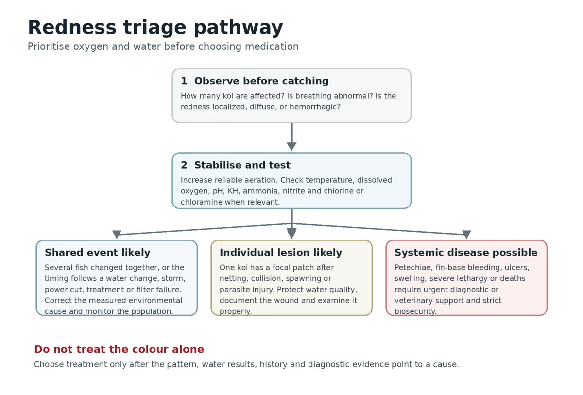
Figure 2 A practical triage pathway for a newly red koi. The sequence protects oxygen and water quality while the cause is narrowed.
How to interpret the first water tests
| Test | Why it belongs in the investigation | Interpretation caution |
|---|---|---|
| Temperature | Changes oxygen capacity, metabolism, ammonia toxicity and drug safety | Use the actual water temperature, not the weather forecast |
| Dissolved oxygen | Low oxygen can cause respiratory distress and magnify every other disease | Measure near the fish and at the riskiest time, often before sunrise |
| pH | Rapid change can injure tissue; pH also changes the toxic ammonia fraction | Compare morning and afternoon and interpret with KH and ammonia |
| KH or alkalinity | Shows buffering reserve and risk of a pH crash | A single pH can look acceptable while buffering is nearly exhausted |
| Total ammonia nitrogen | Signals waste overload or biofilter failure | Toxic NH3 fraction rises with pH and temperature |
| Nitrite | Impairs oxygen transport and indicates incomplete nitrification | Symptoms can resemble low oxygen; chloride protection requires calculation |
| Free and total chlorine | Detects source-water disinfectant exposure | Free chlorine alone can miss chloramine |
| Salinity | Reveals existing salt before any new addition | Evaporation and repeated dosing can leave more salt than expected |
Examining a red koi without making it worse
Plan the examination before netting. Prepare a soft pan net, wet koi sock, adequately sized bowl, air supply, cover, measuring scale, camera and any sampling equipment. Match the bowl water to the pond.
Keep the fish in water and support the body evenly. Avoid dry hands, towels and prolonged restraint.
Observe the fish swimming first. A fish that is upright and responsive may deteriorate after repeated chasing. Record respiratory rate relative to other koi, fin position, balance and response to approach. In the bowl, inspect the eyes, mouth, gill covers, fin bases, vent, belly, scales and every red area. Check for lice, anchor worms, punctures, missing scales, excess mucus and ulcers.
Gill examination and sedation should be performed only by someone competent with koi handling and the chosen anaesthetic. Incorrect concentration, poor oxygen or extended exposure can kill a compromised fish. A brief purposeful procedure is safer than a long improvised one.
When photographing, include one image showing the entire fish so lesion location is clear, then close images with a size reference. Do not alter saturation or apply filters. Record date, water temperature and whether the picture was taken before or after handling.
Diagnostic tests and what they answer
Skin scrape and gill wet mount
A fresh wet mount can demonstrate mobile parasites and show mucus, epithelial cells and debris. It is most valuable when collected from active lesion margins or areas suggested by the clinical signs and examined immediately. Gill samples may be required when breathing is abnormal despite a negative skin scrape.
Microscopy does not prove that every organism seen caused the entire problem. A small parasite burden can coexist with ammonia injury or bacterial disease. Conversely, an apparently negative slide does not eliminate parasites. Sampling, timing and examiner experience affect sensitivity.
Bacterial culture and susceptibility testing
Culture is appropriate when deep ulcers, progressive haemorrhage, systemic signs or deaths suggest bacterial disease. The sample should come from a suitable fresh site using aseptic technique. Surface swabs from old exposed tissue are easily contaminated by environmental bacteria.
Pure isolation and identification help establish significance. Susceptibility testing indicates which antimicrobials inhibit the isolate in the laboratory. The veterinarian still has to consider whether the drug can reach the infected tissue, whether the fish is eating, the formulation available, water temperature, safety and legal restrictions.
PCR histology imaging and necropsy
PCR may be needed when KHV, CEV or another specific infectious agent is a concern. Sample type, temperature history, disease stage and transport conditions influence the result. A negative result does not compensate for a poor sample.
Histology can distinguish inflammation, necrosis, vascular injury, parasites and organ disease that look similar externally. Ultrasound or radiography may help when a single fish has swelling or an internal mass. Necropsy of a freshly dead or appropriately euthanised fish can be the most efficient way to protect the rest of a valuable collection. Coordinate with the receiving laboratory before collecting or freezing specimens.
Treatment principles
Correct the environment first
Environmental correction is treatment when ammonia, chlorine, nitrite, unstable pH, low oxygen or chemical exposure is present. Restore aeration and circulation, remove decaying waste, stop the source, and use conditioned replacement water when indicated. Make changes large enough to reduce the measured danger but controlled enough to avoid a second temperature or chemistry shock.
Keep testing. A value can rebound if the biofilter remains damaged or the contaminant source is still present. Record the time and result before and after each correction. Clear water and calmer fish are encouraging, but they do not replace measurements.
Use quarantine intelligently
Isolation can reduce spread, simplify observation and allow targeted treatment. It can also place the fish into a small uncycled tank with worse oxygen and ammonia than the main pond. The quarantine system needs mature biological capacity, strong aeration, cover, stable temperature and dedicated nets, hoses and test equipment.
Do not share water between quarantine and the main collection. Avoid carrying wet hands, bowls or splash between systems. When several fish in the same pond are affected, moving only the reddest koi may not address the shared cause.
Treat proven parasites and protect wounds
If microscopy or direct observation supports a parasite, select a treatment that covers that organism and is compatible with the system. Calculate volume accurately, check temperature and oxygen requirements, review salt and other recent products, and follow the current legal label. Re-examine when appropriate rather than assuming one dose ended the life cycle.
For a local wound, excellent water and prevention of further trauma are foundational. Professional topical treatment may be suitable for selected superficial lesions. Deep, expanding or necrotic wounds require more than cosmetic cleaning. Excessive scrubbing removes viable tissue and mucus and can enlarge the lesion.
Use antimicrobials only for a justified bacterial case
Antimicrobials do not correct ammonia, chlorine, parasites, viruses or physical injury. Unnecessary use can damage the biofilter, stress fish, contaminate the environment and select resistant bacteria. Human tablets, livestock powders and leftover injections should not be adapted by guesswork.
When systemic bacterial disease is likely, veterinary treatment may involve injection, medicated food or another controlled route. Culture and susceptibility testing are preferable when practical. The treatment plan should include the environmental factors that allowed disease to emerge; otherwise a drug may suppress the infection while the pond continues to injure the fish.
Salt is not a universal redness treatment
Controlled salinity can reduce osmoregulatory work and chloride can protect against nitrite uptake in selected situations. It does not cure every bacterial infection, does not remove every parasite and does not neutralise chlorine. Salt may be incompatible with plants, invertebrates or some medications.
Measure existing salinity, true water volume and the reason for use. Repeated additions without accounting for water removal can create an unexpectedly high concentration. If salt is being considered for nitrite, calculate the chloride requirement from the measured nitrite and obtain appropriate guidance rather than applying a fixed hobby rule to every pond.
Common scenarios
Scenario one Fine tail veins after delivery
A newly imported koi shows fine red lines in the tail after unpacking. It remains upright, breathes normally and becomes interested in food after settling. The other fish are normal and water tests are acceptable.
The history and limited pattern make transport and handling stress plausible. Provide stable oxygen-rich quarantine water, avoid further netting, document the tail and monitor. Do not begin antibiotics merely to make the vessels disappear. Escalate if the redness spreads, appetite falls, breathing changes or a lesion develops.
Scenario two Several koi turn pink after a water change
Within an hour of a large refill, several koi become restless and the white skin looks pink. They breathe faster and gather near returns.
This is a shared environmental emergency until proven otherwise. Increase aeration, test free and total chlorine, compare temperature and pH, confirm dechlorinator delivery and inspect the carbon or dosing system. Correct the measured exposure. Moving one fish or adding an antibacterial treatment would miss the population-level problem.
Scenario three One red patch after spawning
A female has a red patch and two missing scales on one flank after vigorous spawning. She remains active and the other koi have superficial abrasions.
Trauma is likely, but the wound still needs clean water and monitoring. Inspect the pond for rough surfaces, document lesion size, and minimise additional handling. If the patch expands, becomes raised, loses tissue or the fish stops feeding, examine for secondary infection and seek targeted care.
Scenario four Red veins with flashing
Several koi flash, clamp their fins and show more visible vessels. Ammonia and nitrite are undetectable, but a new batch arrived two weeks earlier.
Parasites become a meaningful differential, but the signs are not specific. Review pH, chlorine and treatment history, then obtain fresh skin and gill samples. Treat the organism found rather than rotating through general products. Keep equipment separate from unaffected systems.
Scenario five Petechiae ulcers and rapid decline
A koi develops pinpoint haemorrhages, red fin bases, two ulcers, pop-eye and severe lethargy. A second fish stops eating.
This pattern is compatible with serious systemic disease and demands urgent support. Stabilise oxygen and water, stop transfers, use biosecurity and contact an aquatic veterinarian or diagnostic laboratory. Culture, susceptibility testing, microscopy, PCR or necropsy may be required. A random pond antibiotic can delay the correct diagnosis and expose unaffected fish.
Scenario six Redness after medication
Fish become red and breathe heavily after an oxidising treatment. The pond contains a high organic load and aeration is marginal.
Assume treatment-related irritation or oxygen stress is possible. Stop adding products, increase aeration, check the label's emergency guidance, measure water parameters and contact the manufacturer or a fish-health professional if signs persist. Do not neutralise one chemical with another unless the interaction is known and specifically advised.
Malaysia and warm climate considerations
Malaysian ponds often operate at warm temperatures with year-round feeding. This can support growth but narrows the oxygen reserve, particularly in heavily stocked dealer systems. Bacterial growth and parasite life cycles may also progress quickly. A red koi in warm water should therefore be assessed promptly rather than watched for a week without measurements.
Heavy rain can dilute alkalinity, move pH, cool the surface, wash contaminants or organic material into an open pond and alter stratification. The effect depends on roof runoff, pond design and source water. Test after major storms. Do not assume rain automatically improves oxygen or that clear rainwater is chemically neutral.
Frequent import, auction and consolidated-shipping activity increases the importance of true quarantine. Fish from different sources should not share water, nets, bowls or wet hands until the health status is understood. A visible red vein may be minor, but mixing groups before investigation can turn an uncertain individual case into a business-wide biosecurity problem.
For valuable koi, recurrent unexplained disease or multiple deaths, contact an aquatic veterinarian or a diagnostic laboratory before collecting samples. The Department of Fisheries Malaysia publishes a current list of fisheries biosecurity laboratories. Confirm submission requirements, sample containers, temperature control and forms before transport. [7]
Prevention and operating discipline
- Maintain enough aeration for the hottest weather, the highest feeding period and a pump failure, not merely for an average day.
- Record temperature, pH, KH, ammonia, nitrite and unusual oxygen readings so change is visible before fish become red.
- Verify dechlorination with testing. Service carbon and dosing equipment according to measured throughput, not memory.
- Remove solids promptly and keep mechanical stages easy to inspect. Organic accumulation consumes oxygen and supports opportunistic bacteria.
- Quarantine new koi in a genuinely separate system with dedicated equipment and adequate biological filtration.
- Photograph valuable koi while healthy. Baseline images make later vessel and skin changes easier to judge.
- Use soft, wet handling equipment and train every worker in netting, sock transfer, bowl oxygen and record keeping.
- Inspect pond surfaces and transport boxes for sharp or abrasive points. Fix repeated injury sources instead of repeatedly treating wounds.
- Do not combine treatments without a documented compatibility check. Keep a log of product, active ingredient, amount, actual volume, time and response.
- Establish a relationship with a fish-health professional and laboratory before an outbreak, when there is time to learn sample requirements.
Daily monitoring checklist
- Is the redness less, unchanged or spreading under the same light and viewing angle?
- Are fine vessels becoming diffuse patches, petechiae, fin-base bleeding or an ulcer?
- Is the koi upright and swimming with the group?
- Is breathing calm compared with unaffected fish?
- Is appetite returning, stable or declining?
- Are fins open or clamped? Is the fish flashing, jumping or isolating?
- Are scales missing, raised or surrounded by swelling?
- Are eyes, abdomen and vent normal?
- Have any other koi developed colour, breathing or behaviour changes?
- What are today's temperature, oxygen, pH, KH, ammonia, nitrite, chlorine and salinity results where relevant?
- What actions were taken, and what changed after each action?
Common mistakes
Calling every red vein septicaemia
Fine visible vessels can occur with stress or local irritation. Calling every case septicaemia leads to unnecessary antibiotics and distracts from water, oxygen and handling.
Assuming stress explains everything
Stress is a physiological state and a risk factor, not a complete diagnosis. If redness progresses, petechiae appear or appetite and breathing worsen, investigate rather than waiting for the fish to settle.
Treating before testing
A medication can alter microscopy, damage gills, lower oxygen or injure the biofilter. Obtain the most useful water and diagnostic evidence first unless an immediate environmental correction is required.
Moving the fish into an unsafe hospital tank
A small uncycled tank can produce ammonia and oxygen problems within hours. Isolation should improve control and biosecurity, not create a second emergency.
Using water clarity as proof of safety
Chlorine, ammonia, nitrite, low oxygen and pH instability can exist in clear water. Measurements and fish behaviour matter more than appearance.
Mixing salt oxidisers and parasite treatments
Compatibility depends on the exact chemicals, concentration, organic load, temperature and fish condition. A succession of products can create more redness while obscuring the original cause.
Photographing with heavy filters
Automatic enhancement, warm lighting and high saturation distort red tissue. Use consistent light and preserve original files so progress can be judged honestly.
Frequently asked questions
Are red veins always bacterial
No. They can become visible with handling, heat, low oxygen, chemical irritation, water-quality stress, parasites or local injury. Bacterial disease becomes more concerning when haemorrhage is widespread or accompanied by ulcers, swelling, anorexia, lethargy or deaths.
Can stress alone make a koi look red
Stress can alter circulation and make vessels more obvious, particularly on white skin and fins. The word stress should still lead to a search for the stressor, such as oxygen loss, transport, temperature change, crowding or poor water.
Should I add salt immediately
Not automatically. Salt has specific supportive uses, including selected nitrite and osmoregulatory situations, but it is not a universal treatment for redness. Measure salinity, volume and the actual problem first.
Should I use an antibiotic food
Only when bacterial disease is reasonably supported and the fish is eating enough to receive a calculated dose. Culture and susceptibility testing are preferable. A fish that refuses food cannot be treated reliably through feed.
Can ammonia cause red veins
Ammonia can injure gills and skin and create irritation, abnormal breathing and colour change. Test total ammonia and interpret it with pH and temperature. Do not rely on redness to estimate severity.
Why are only my white koi showing redness
The contrast makes vascular change easier to see. Dark or patterned koi may be affected without looking equally red. Compare behaviour and water exposure across the whole group.
How long should transport redness take to settle
There is no universal safe time. A mild vessel change should trend down as the fish stabilises. Spreading redness, abnormal breathing, poor appetite or new lesions are reasons to investigate immediately rather than wait for a fixed number of days.
Is redness contagious
Redness itself is not contagious. Its cause may be shared or infectious. Several affected fish, a new introduction, gill disease or deaths justify biosecurity until the cause is clearer.
When should I take a skin scrape
When flashing, excess mucus, clamped fins, new introductions or an unexplained group problem make parasites plausible. Gill sampling may be needed when respiratory signs dominate.
When should I contact a veterinarian or laboratory
Promptly when there are petechiae, deep ulcers, swelling, pop-eye, severe breathing difficulty, rapid deterioration, several affected koi or unexplained deaths. Early fresh samples usually provide more information than late decomposed material.
Conclusion
Red veins and reddening are among the most useful but most easily overinterpreted signs in koi. They can be the temporary visible effect of handling, the first evidence of a water-quality failure, the margin of a local wound, the result of parasite irritation or part of a life-threatening haemorrhagic disease. The colour alone cannot decide which explanation is correct.
The reliable approach is disciplined: observe before catching, count affected fish, support oxygen, test the water, reconstruct the recent history, document the pattern and choose diagnostics that answer the next question. Correct environmental causes immediately. Use microscopy for plausible parasites, culture and susceptibility testing for serious bacterial disease, and PCR or necropsy when the population pattern demands it.
The aim is not simply to make the red colour disappear. It is to identify and remove the process that made the tissue red while protecting the rest of the collection from the same event.
References and further reading
This article is an original educational synthesis. It is intended to help koi keepers organise observations and seek appropriate support; it does not replace examination of an individual fish by a qualified aquatic veterinarian or diagnostic laboratory. Product labels, permitted uses and regulations can change. Check current local requirements before using any treatment.
1. Petty BD, Francis-Floyd R, Yanong RPE. Bacterial Diseases of Fish. Merck Veterinary Manual. Full review May 2022; updated September 2024. https://www.merckvetmanual.com/exotic-and-laboratory-animals/aquarium-fish/bacterial-diseases-of-fish
2. Francis-Floyd R, Petty BD. Routine Health Care of Fish. MSD Veterinary Manual. Full review February 2020; updated April 2026. https://www.msdvetmanual.com/all-other-pets/fish/routine-health-care-of-fish
3. Francis-Floyd R, Petty BD. Emergencies of Fish. Merck Veterinary Manual. Updated September 2024. https://www.merckvetmanual.com/all-other-pets/fish/emergencies-of-fish
4. Francis-Floyd R. Stress Its Role in Fish Disease. University of Florida IFAS Extension FA005. https://ask.ifas.ufl.edu/publication/FA005
5. Yanong RPE. Fish Health Management Considerations in Recirculating Aquaculture Systems Part 3 General Recommendations and Problem Solving Approaches. University of Florida IFAS Extension FA101. https://ask.ifas.ufl.edu/publication/FA101
6. Petty D, Hartman KH, Yanong RPE, Pouder DB, Francis-Floyd R, Waltzek TB. Koi Herpesvirus Disease. University of Florida IFAS Extension VM113. https://ask.ifas.ufl.edu/publication/VM113
7. Department of Fisheries Malaysia. List of Fisheries Biosecurity Laboratories. https://www.dof.gov.my/en/services/certification-scheme/list-of-fisheries-biosecurity-laboratories/
8. Noga EJ. Fish Disease Diagnosis and Treatment. 2nd edition. Wiley Blackwell. 2010. https://www.wiley.com/en-us/Fish+Disease%3A+Diagnosis+and+Treatment%2C+2nd+Edition-p-9781119949466
9. Roberts RJ, editor. Fish Pathology. 4th edition. Wiley Blackwell. 2012. https://www.wiley.com/en-us/Fish+Pathology%2C+4th+Edition-p-9781444332827
10. Stoskopf MK, editor. Fish Medicine. W B Saunders. 1993. https://www.worldcat.org/title/26764321
11. Austin B, Austin DA. Bacterial Fish Pathogens Disease of Farmed and Wild Fish. 6th edition. Springer. 2016. https://link.springer.com/book/10.1007/978-3-319-32674-0
12. Wedemeyer GA. Physiology of Fish in Intensive Culture Systems. Springer. 1996. https://link.springer.com/book/10.1007/978-1-4615-6011-1
13. Evans DH. Teleost fish osmoregulation what have we learned since August Krogh Homer Smith and Ancel Keys. American Journal of Physiology. 2008;295:R704-R713. https://pubmed.ncbi.nlm.nih.gov/18550624/
14. Greenwell MG, Sherrill J, Clayton LA. Osmoregulation in fish mechanisms and clinical implications. Veterinary Clinics of North America Exotic Animal Practice. 2003;6:169-189. https://pubmed.ncbi.nlm.nih.gov/12616840/
15. World Organisation for Animal Health. Infection with koi herpesvirus. Aquatic Manual and disease information. https://www.woah.org/en/disease/koi-herpesvirus-disease/
16. Timmons MB, Ebeling JM. Recirculating Aquaculture. 3rd edition. Ithaca Publishing Company. 2013. https://www.worldcat.org/title/853447500
17. Emmenegger E. Koi infected with spring viraemia of carp virus. U.S. Geological Survey. Public domain photograph. https://www.usgs.gov/media/images/investigation-and-disease-prevention-spring-viremia-carp-virus
18. Laslovarga. Koi Fish pond. Wikimedia Commons. CC BY-SA 3.0 photograph. https://commons.wikimedia.org/wiki/File:1Koi_Fish.JPG